Traditional diagnosis
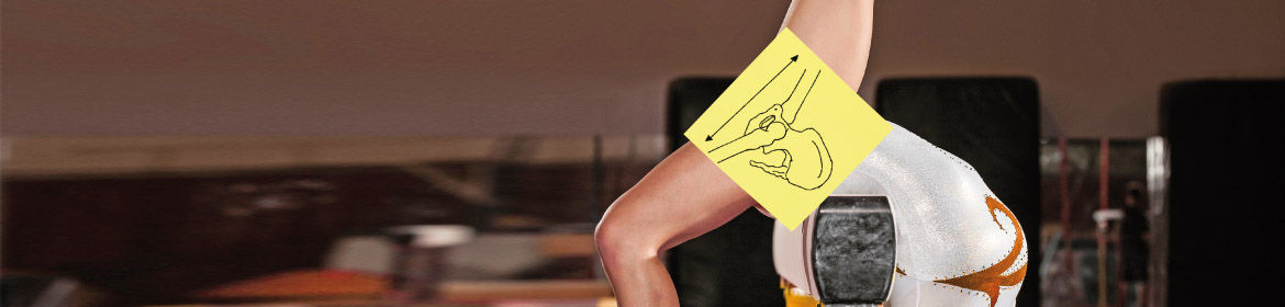
The sooner FAI is detected, the better. However, early stage FAI can be difficult to detect. Pain complaints may be caused by a variety of reasons, so radiological images are required to inspect the joint. Every surgeon will have a personal preference on the type of diagnostic images to use for diagnosis and assessment of FAI. Some choose MR (based on magnetism), some choose CT (based on x-rays), and some only use simple radiographs.
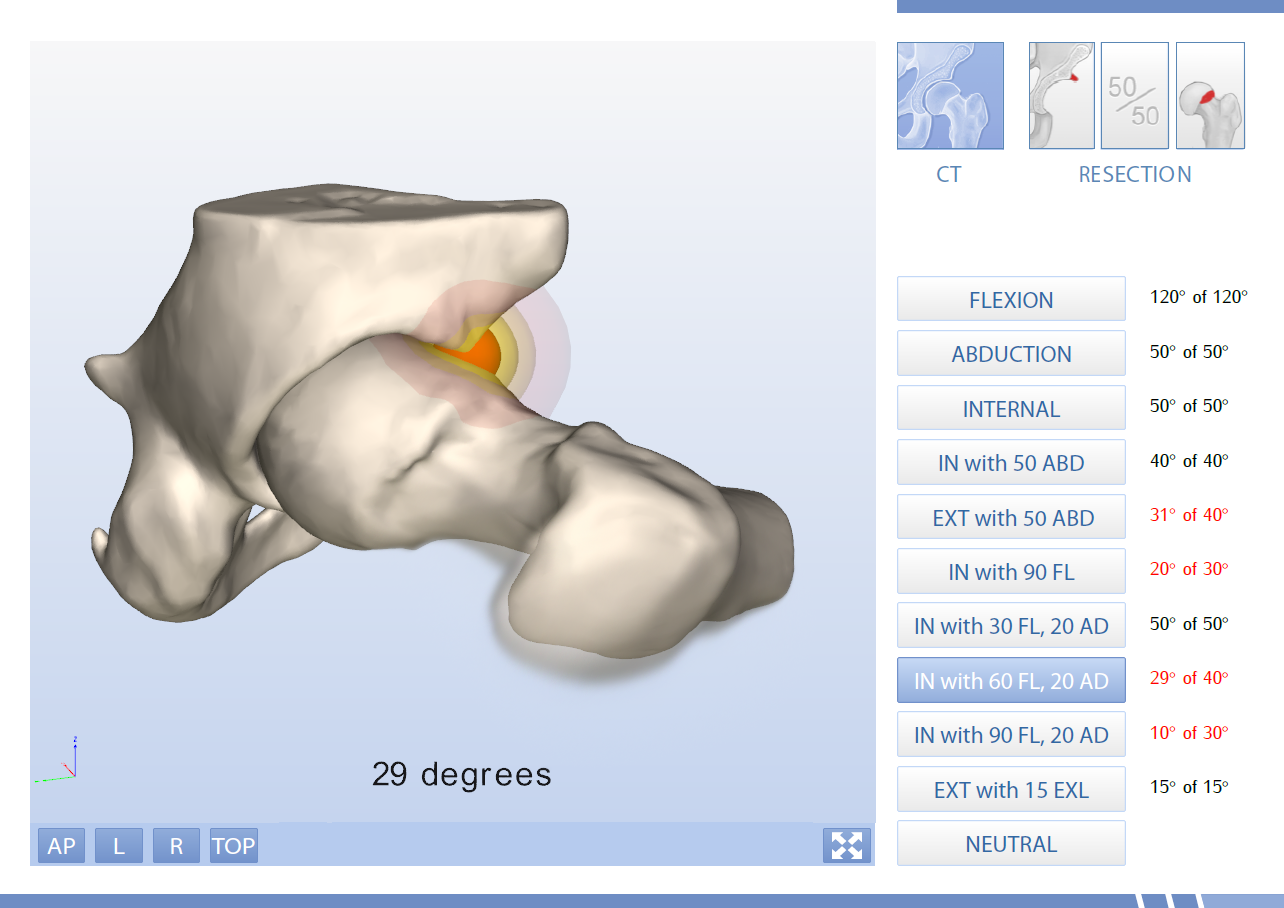
Not every shape deviation in the hip joint will be a cause for hip impingement. Therefore, the challenge is to determine which shape deviations are causing hip impingement.
About the Move Forward™ 3D motion simulation service of Clinical Graphics
At Clinical Graphics we provide the Move Forward™ service. We automatically analyze CT and MRI scans, then produce interactive 3D PDF reports that will tell you whether the shape of a joint is likely to cause FAI. If this is the case, the software also calculates what should be done to resolve hip impingement, alleviate pain and increase the functional range of motion of the hip joint.