
What is hip impingement? What is femoroacetabular impingement (FAI)?
Femoroacetabular impingement (FAI), also known as hip impingement, is a condition where the bones of the hip joint do not have a matching shape. During movement this mismatch leads to friction in the joint. Depending on the severity of the mismatch and the activities this may damage the cartilage and tissues surrounding the joint. The symptoms of FAI are pain of the hip and a reduced range of motion. When the cup gives too much coverage, leading to impingement, this is called Pincer type impingement. When the head of the femur has a bump that causes impingement, this is called a Cam type impingement.
Hip arthroscopy, a rising star
In many cases of hip impingement it is possible to perform 'joint preserving surgery'; the surgeon removes the excess bone and tries to maintain as much of your hip joint as possible. When the bone areas that caused impingement are removed, the joint can move freely again. Joint preserving surgery may allow you to return to practicing active sports again. An increasing number of surgeons perform this type of surgery arthroscopically: they use a camera and tools for which no incision is required. The advantage of an arthroscopic procedure compared to open surgery is that less damage is being done to the tissues surrounding the joint.
Traditional diagnosis for FAI, hip impingement
The sooner hip impingement is detected, the better. However, early stage FAI can be difficult to detect. Pain complaints may be caused by a variety of reasons, so radiological images are required to inspect the joint. Every surgeon will have a personal preference on the type of diagnostic images to use for diagnosis and assessment of hip impingement. Some choose MR (based on magnetism), some choose CT (based on x-rays), and some only use simple radiographs. Not every shape deviation in the hip joint will be a cause for hip impingement. Therefore, the challenge is to determine which shape deviations are causing hip impingement and should therefore be removed in a surgical procedure.
Today's static imaging leaves much to the imagination
when it comes to diagnose femoroacetabular impingement (FAI) or hip impingement.
Our Move Forward™ 3D motion simulations for hip impingement based on CT or MRI exams are the missing link and enable you to see beyond & to be sure.
Already want to request a report?
Move Forward™ then!
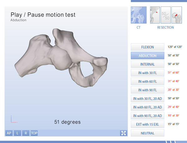
Vizualising hip impingement or FAI: an interactive 3D view in motion from all angles
The bone shape variations that cause hip impingement are sometimes missed with traditional radiology. The complex relationship between the shape of the cup, the shape of the femoral head and how these interact during motion is difficult to picture without an actual visual representation in 3D motion. Joint problems are dynamic and multi dimensional. Today's static imaging leaves much to the imagination when it comes to diagnose hip impingement. Our Move Forward™ 3D motion simulations for hip impingement, femoroacetabular impingement or short FAI, are the missing link. Our 3D motion simulation reports provide an interactive 3D view of the hip joint in motion from all angles and show the exact point of impingement within the hip joint, based on patient specific traditional CT or MRI exams.
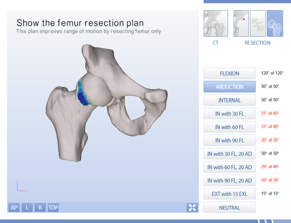
Visualizing surgery plans and potential effects of treatment
If femoracetabular impingement (FAI) or hip impingement is detected and range of motion is limited, simulated surgery plans that each individually restore the range of motion of the hip joint and the potential effects of the operation plans are included in the 3D motion analysis for hip impingement as well. In addition to these advanced calculations, the reports include radiographic parameters such as acetabular version, joint coverage, alpha angles and LCE angles. Clinical judgment completes the equation to optimize clinical outcome.
 Are you a medical professional? Get your free trial now!
Across the world many medical professionals already use our hurdle free Move Forward™ 3D motion simulation service for hip impingement. It would be our pleasure to show you the value of our service too. We invite you to request a free trial! No strings attached.
 Are you a patient? See beyond & move forward too! See your case in 3D motion
Our Move Forward™ 3D motion simulation reports for hip impingement are delivered to medical professionals only. If you are interested to get a report of your case, either contact a connected medical professional or request a free brochure to inform your doctor about the Move Forward™ 3D motion simulation reports.